
ขอแสดงความยินดีกับทีมวิจัย ผศ.ดร.ป๋วย อุ่นใจ จากภาควิชาชีววิทยา และ ศ.ดร.เจมส์ เกตุทัต คาร์นส์ มหาวิทยาลัยเทคโนโลยีสุรนารี สำหรับผลงานการค้นพบโครงสร้างของเอนไซม์ GH116 จากพืช (AtGCD3) ด้วยเทคนิค Cryo-Electron Microscopy (Cryo-EM)
ขอแสดงความยินดีกับทีมวิจัย ผศ ดร ป๋วย อุ่นใจ จากภาควิชาชีววิทยา คณะวิทยาศาสตร์ มหาวิทยาลัยมหิดล และ ศ ดร เจมส์ เกตุทัต คาร์นส์ มหาวิทยาลัยเทคโนโลยีสุรนารี สำหรับผลงานการค้นพบโครงสร้างของเอนไซม์ GH116 จากพืช (AtGCD3) ด้วยเทคนิค Cryo-Electron Microscopy (Cryo-EM) ซึ่งนับเป็นโครงสร้างของเอนไซม์กลุ่ม GH116 จากสิ่งมีชีวิตยูคาริโอตที่ได้รับการอธิบายเป็นครั้งแรก ความสำเร็จครั้งนี้ช่วยเปิดมุมมองใหม่ต่อความเข้าใจเกี่ยวกับเอนไซม์ที่ทำหน้าที่ย่อย glucosylceramide (GlcCer) ซึ่งเป็นไขมันชีวโมเลกุลสำคัญที่เกี่ยวข้องกับกระบวนการทางชีวภาพทั้งในพืชและสัตว์
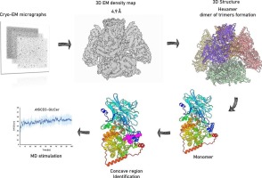
จากการศึกษาพบว่าโปรตีน AtGCD3 มีโครงสร้างเป็น hexamer ที่ประกอบด้วยการรวมตัวของ trimer สองชุด โดยมีแรงยึดเหนี่ยวทั้งแบบ hydrophobic interactions, hydrogen bonds และ salt bridges ช่วยเสริมความเสถียรของโครงสร้าง นอกจากนี้ยังพบองค์ประกอบโครงสร้างใหม่คือ α-helix สองเส้น ที่ปกคลุมบริเวณ active site และก่อให้เกิดช่องทางไฮโดรโฟบิกซึ่งอาจมีบทบาทสำคัญในการจับกับส่วน lipid tail ของ glucosylceramide
การจำลองเชิงคำนวณด้วย molecular dynamics simulation ยังสนับสนุนว่าโมเลกุล GlcCer สามารถจับกับเอนไซม์ได้อย่างเสถียร โดยส่วนของไขมันจะวางตัวอยู่ภายในช่องทางไฮโดรโฟบิกดังกล่าว การค้นพบนี้ไม่เพียงช่วยอธิบายกลไกการทำงานของเอนไซม์ GH116 ในพืชเท่านั้น แต่ยังมีความสำคัญต่อการสร้างแบบจำลองโครงสร้างของเอนไซม์ GBA2 ในมนุษย์ ซึ่งเกี่ยวข้องกับโรคสำคัญ เช่น Gaucher disease และ hereditary spastic paraplegia
ผลงานวิจัยนี้จึงถือเป็นอีกก้าวสำคัญของวงการชีววิทยาโครงสร้าง ที่เชื่อมโยงองค์ความรู้จากพืชสู่การทำความเข้าใจโรคในมนุษย์ ขอชื่นชมและแสดงความยินดีกับนักวิจัยทุกท่านสำหรับความมุ่งมั่น ความพยายาม และการสร้างองค์ความรู้ใหม่ที่มีคุณค่าอย่างยิ่งต่อวงการวิทยาศาสตร์และการแพทย์ในอนาคต 
งานนี้มีนักวิจัยจากจุฬาลงกรณ์มหาวิทยาลัย VISTEC และ MUFRF ร่วมด้วย
แต่ที่น่าตื่นเต้น ก็คือ โครงสร้างนี้ คือโครงสร้างแรกจาก State of the Art CryoEM จากประเทศไทย!!!!
Congratulations to the research team led by Asst. Prof. Dr. Puey Ounjai from the Department of Biology, Faculty of Science, Mahidol University, and Prof. Dr. James Ketudat Cairns from Suranaree University of Technology for their remarkable discovery of the structure of the plant GH116 enzyme (AtGCD3) using Cryo-Electron Microscopy (Cryo-EM). This represents the first reported structure of a GH116 enzyme from a eukaryotic organism. The achievement provides new insights into enzymes responsible for degrading glucosylceramide (GlcCer), an important biomolecular lipid involved in biological processes in both plants and animals.
The study revealed that the AtGCD3 protein forms a hexameric structure, composed of two trimers. The assembly is stabilized by hydrophobic interactions, hydrogen bonds, and salt bridges. In addition, the researchers identified two α-helices not previously observed in other GH116 structures. These helices cover the active site and create hydrophobic channels that may accommodate the lipid tails of glucosylceramide.
Computational analysis using molecular dynamics simulations further supports that GlcCer can bind stably within the enzyme’s active site, with its lipid tails positioned in these hydrophobic channels. This discovery not only advances our understanding of the catalytic mechanism of GH116 enzymes in plants but also provides a valuable structural template for modeling the human enzyme GBA2, which is associated with diseases such as Gaucher disease and hereditary spastic paraplegia.
This research marks an important milestone in structural biology, linking knowledge from plant systems to the understanding of human disease mechanisms. We commend the dedication and achievements of all researchers involved.
The work also includes contributions from researchers at Chulalongkorn University, VISTEC and MUFRF.
Perhaps most exciting of all, this structure represents the first structure determined using state-of-the-art Cryo-EM technology in Thailand.